Getting a clearer picture of brain imaging
Emory technology BrICS aids doctors in making informed decisions for brain cancer patients
Traditional brain imaging like CT scans or MRIs can help us see our brains, including its structure and diseases like tumors. However, they cannot see subtle changes in brain metabolism and often cannot differentiate between different types of brain injury, such as recurrence of a brain tumor or the damage that can occur from radiation therapy. Traditionally, finding out these kinds of details would require a surgical procedure to sample a piece of brain tissue.
Using a newer technology called spectroscopic MRI (sMRI), developed by Hyunsuk Shim, PhD and her team at Emory University and the University of Miami, physicians and researchers can get more specific information about the chemical makeup of different areas of the brain, using it to map out tumors and guide treatment like radiation to problematic regions undetected in standard imaging. Even though sMRI can help see differences in brain tissue, the data is complex and can be hard for doctors to see them in a way that helps them make decisions. That’s where a new technology, BrICS, the Brain Imaging Collaborative Suite, comes into play. BrICS is web-based software doctors can use to review whole-brain spectroscopic MRIs and make appropriate decisions for treating their patients.

“There's a huge metabolic difference between tumor cells and surrounding normal tissues in the brain, so by using that, we can tell the difference between areas of tumor and normal brain,” Shim explained. “Radiation oncologists and neuroradiologists can then use this to identify the tumor better than standard imaging.”
BrICS displays sMRI results by overlaying color-coded metabolic ratio maps on anatomical brain images.
“With BrICS, we provide features for doctors to see areas of the brain tumor that they couldn't see before, and we also provide tools for them to target those areas where there is metabolic activity that's really abnormal,” said Karthik Ramesh, a developer of BrICS, who is a doctoral student on Shim’s team.
Shim’s team has used the product in a multisite clinical trial that successfully treated 30 patients, showing a promising improvement in survival outcomes. Now, Shim and her team hope to disseminate the technology to more hospitals to be used in everyday clinical workflows.
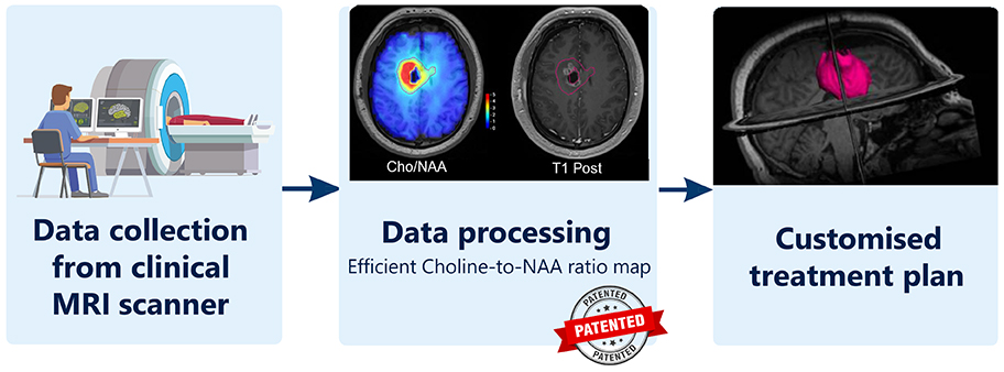
Schematic of the BrICS platform
“It is exciting to see how sMRI is going to change the way we view and treat glioblastoma. I am looking forward to working with Dr. Shim to get the technology out to the marketplace,” expressed Catherine Murari-Kanti, a senior licensing associate in Emory’s Office of Technology Transfer. “These technologies that improve the lifespan of cancer patients speak to the very essence of technology transfer.”
The journey for BrICS is far from over. The second step is refining and spreading this new tool, Shim explained. “We want to put our technology on MR scanners everywhere which will expedite processing of image results and allow clinicians to view our imaging immediately.”
The most gratifying part for Shim and her team is making a difference for patients. “We actually made patient outcomes different using this imaging compared to the previous standard outcome,” Shim said.
— Chaya Tong
Techid: 18245
Read our technology brief